Special Exhibit: Early American Photomicrographs by Joseph Janvier Woodward
Click here to view the Exhibition Images
In 1862, during the US Civil War, the medical officer Joseph Janvier Woodward (1833-1884) was selected for the US War Department's newly-founded medical research facility, the Army Medical Museum. Woodward's duties included management of the Museum's medical work and microscopy as well as production of the three medical volumes for the massive Medical and Surgical History of the War of the Rebellion (1870-1888) whose final volume was published four years after his death. (Burns, 1983, p.1463)
In 1869 Woodward became the first microscopist to successfully resolve the 19th band of Norbert's 19-band test plate, created as test object for the resolution of period microscopes. While this was not understood at the time, Woodward was thereby approaching the absolute limits of the optical microscope, and the subsequent 20-band test-plates produced by Norbert contained rulings only capable of resolution by methods such as the electron microscope. (Turner, 1980, p.170)
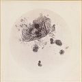
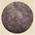
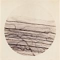
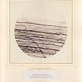
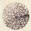
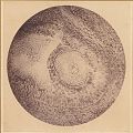
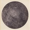
Woodward's work at the Army Medical Museum involved extensive documentation through microphotography where again his work was on the frontier of period science. At the Museum he implemented important advances in microscopes, light-sources, tissue-dying techniques and integration of photography with microscopy, turning America into a world-leader in microphotography (Burns, 1983, p.1463). Woodward has been called "the most skillful microphotographer of his age" (Frison 1954, p.120).
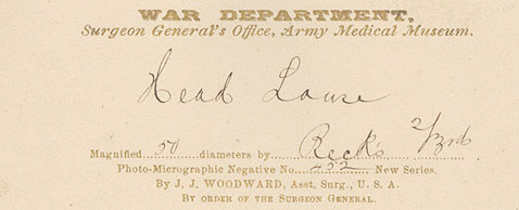
Woodward published several pamplets illustrating his advances with the microscope, often accompanied by superb 6 x 6 inch (16x16 cm) albumen prints of his photomicrographs on special gilt-bordered US War Department mounts. These typically measure 14 x 11 inches (35x28 cm) and bear the imprint "War Department, Surgeon General's Office, Army Medical Museum" ... "By J. J. Woodward".
Nine such plates were intended to accompany his Report to the Surgeon General of the United States Army on an Improved Method of Photographing Histological Preparations by Sunlight (1871), The plates bear pasted-on print labels identifying each subject as well as its magnification, series number, and other notations.
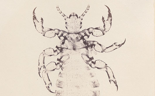
Woodward's Army Medical Museum plates were also issued in reference volumes and were presumably available to researchers individually. Thus, Woodward's photomicrograph of a Head Louse is found on his standard gilt War Department mount but with manuscript notations of subject and magnification rather than a printed label, perhaps indicating that it was issued one-off and not as part of a series.
Woodwards beautiful microscopic images present a jarring intersection between the onset of advanced scientific technology and an era we more often associate with muskets and hardtack.
Images and text copyright © 2006 by Christopher Wahren Fine Photographs
Click here to view the Exhibition Images ![]()
...or here to view J. J. Woodward photographs offered in the Sales Galleries
![]()
Works Cited & Recommended Reading:
Woodward, Joseph Janvier, Report to the Surgeon General of the United States Army on an Improved Method of Photographing Histological Preparations by Sunlight. Washington D.C, 1871.
Frison, Ed., L'Evolution de la Partie Optique de Microscope au cours du Dix-Neuvieme Siecle. Leyde, 1954.
Turner, G. L'E., Essays on the History of the Microscope. Oxford, Senecio Publishing, 1980.
Burns, Stanley, Early Medical Photography in America (1839-1883). New York, The Burns Archive, 1983.
Links:
Woodward and Medicine in Archives of Pathology & Laboratory Medicine, Oct 2005
On Woodward and The Medical and Surgical History of the Rebellion